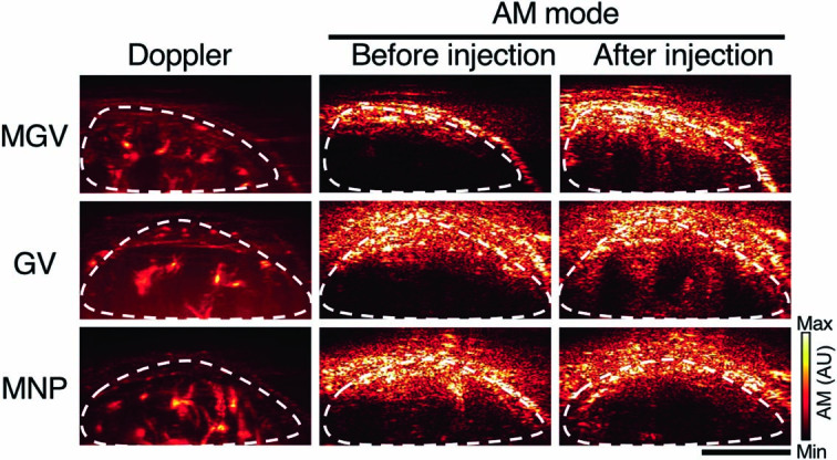
Extended Data Fig. 7. Tracking GV and MGV delivery using AM and Doppler imaging in the liver.

Three different groups of nanomaterials (MGVs, GVs, or MNPs) were injected intravenously into different cohorts, and liver AM and Doppler images were taken after 5 minutes. Doppler images reveal the position of the liver, AM images show the signal from GVs or MGVs after injection (n = 3 animals per group). Scale bars, 5 mm. Min and max on colour bar ranges from 0 to 1000000 arbitrary units.
