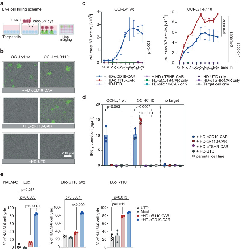
Fig. 3. Humanization of the binding moiety of chimeric antigen receptor (CAR) T cells against the IGLV3-21R110 neoepitope.
a Schematic representation of the applied live cell killing assay using the Incucyte system and the Caspase-3/7 fluorescence dye. Created with BioRender.com. b–d Cytolysis of IGLV3-21R110 expressing OCI-Ly1-R110 cells and OCI-Ly1 wild-type (wt) cells by healthy donor anti-IGLV3-21R110 CAR T cells with a humanized scFv sequence (HD-αR110-CAR) with an E:T ratio of 5:1. Cytolysis was monitored over time as caspase 3/7 activity (relative intensity of green fluorescence) in co-cultures using Incucyte S3 (c). Anti-thyroid stimulating hormone receptor (TSHR) CAR T cells (HD-αTSHR-CAR) and untransduced T cells (UTD) served as controls. All conditions indicated have been plotted, negative control conditions overlap. b Representative images show CAR T cell-mediated cytolysis after 24 h. d Quantification of IFN-γ in co-culture supernatants after 24 h incubation of indicated target/effector cell combinations. e Percent tumor cell lysis based on a bioluminescence assay after 48 h co-culture of NALM-6 Luc expressing IGLV3-21R110 or its non-pathogenic counterpart IGLV3-21G110 in an effector to target (E:T) ratio of 5:1. All bar plots represent the indicated mean ± SD (n = 3) with n indicates independent experiments/donors. Statistics: paired, one-sided t test (c), one-sided t test (d), one-way ANOVA followed by t test with Welch’s correction (e). Source data are provided as a Source Data file.

