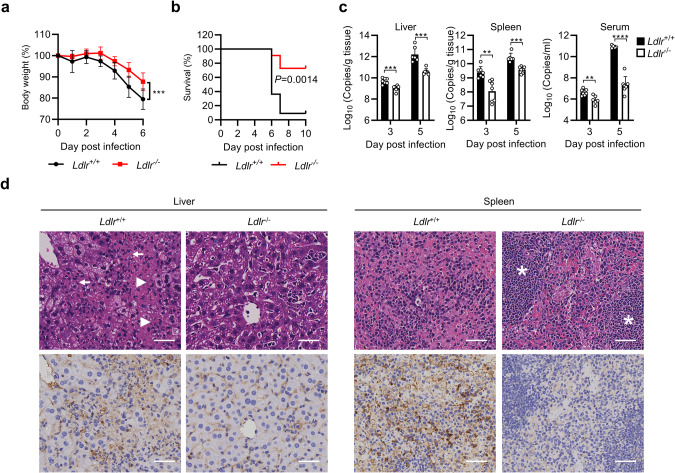
Fig. 5. LDLR is required for CCHFV pathogenesis in mice.
a, b WT (n = 11) and LDLR-knockout (n = 11) mice were pretreated with anti-IFNAR1 monoclonal antibody MAR1-5A3 (200 µg/mouse) 24 h before infection and then infected with CCHFV (10 TCID50) via the intraperitoneal route. Forty-eight hours post infection, 200 µg of MAR1-5A3 was administrated. The body weight (a) and survival (b) of the mice were monitored daily. c, d WT (n = 7) and LDLR-knockout (n = 7) mice were pretreated with anti-IFNAR1 monoclonal antibody MAR1-5A3 (200 µg/mouse) 24 h before infection and then infected with CCHFV (10 TCID50) via the intraperitoneal route. Forty-eight hours post infection, 200 µg of MAR1-5A3 was administrated. Mice were necropsied and the livers and spleens were collected at day 3 and 5 post infection. Viral loads were quantified by RT-qPCR and shown as the number of viral RNA copies per microgram of organs or per mL of sera (c). H&E staining and immunostaining with anti-Gn mAb (7A11) were performed and the pathological changes were indicated (d). The extensive necrosis (white arrowheads) and necrotic cellular debris (white arrows) in the liver were indicated; white pulps in the spleen (white asterisks) were marked. The bars represent 100 µm. Data are represented as mean ± SD. **P < 0.01; ***P < 0.001; ****P < 0.0001.

