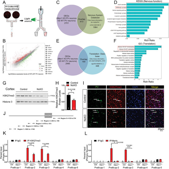
Figure 3.

Transcriptome analysis of Men1 −/− PV neurons revealed epigenetics regulation of pvalb by Menin. A) PcKO mice were crossed with Ai14 tomato reporter mice, and fluorescent PV cells from the brain were subjected for flow cytometry sorting. B–F) Transcriptome sequencing of Men1 knockout and WT PV neurons. Differentially expressed genes (DEGs) were identified and were shown in panel (B). C–F) These DEGs were compared with the nervous system database or the translation database of mouse genome information, and the overlapped genes were subjected to KEGG pathway analysis or GO pathway analysis. G,H) Western blot analysis of H3K27me3 protein expression in primary neuron from NcKO and control mice. Quantification of protein levels is shown in (H), n = 8 mice. (I) Double immunofluorescence staining to detect PV (green) and H3K27me3 (red) in NcKO and control mice cortex. Scale bar:20 µm, n = 3 mice. J) Schematic diagram of pvalb promoter region. K,L) ChIP assays using antibodies against H3K27me3 (K) or Menin (L) were performed in cultured Men1‐knockout or WT neurons on DIV 12. n = 4 independent experiments.Data represent mean±SEM. ns: not significant, * p < 0.05, ** p < 0.01, *** p < 0.001, one‐way ANOVA with Tukey's post hoc analysis. See also Figure S10 (Supporting Information).
