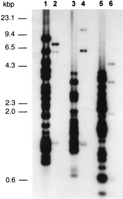
FIG. 5.
Southern blot of genomic DNAs from M. synoviae WVU (lanes 1, 3, and 5) and M. gallisepticum S6 DNA (lanes 2, 4, and 6) digested with restriction enzymes BglII (lanes 1 and 2), EcoRI (lanes 3 and 4), and HindIII (lanes 5 and 6) and probed with the 32P-labelled vlhA gene. The blot was washed under low-stringency conditions and autoradiographed. Three fragments of M. gallisepticum digested with each restriction endonuclease (BglII, 7.4, 5.8, and 1.2 kb; EcoRI, 11.2, 6, and 1.1 kb; and HindIII, 4.5, 3.2, and 1.5 kb) hybridized to the vlhA gene probe. Numbers on the left indicate the sizes of the nucleic acid molecular size markers (HindIII-digested λ phage).

