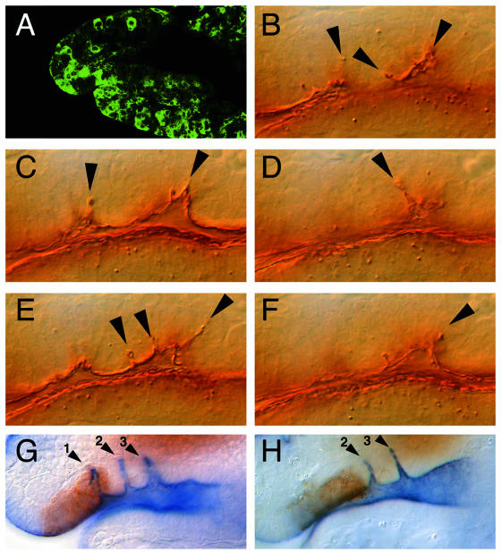
Fig. 3. Expression of secreted Spitz and DERDN at the invaginating SNS. (A) GFP immunolabeling to show embryonic expression of GFP under the control of an actin promotor during stage 11. Genotype Actin-Gal4/UAS-GFP. At this stage, the actin promotor in the Gal4 driver activates scattered embryonic expression of the reporter gene. (B–F) Crumbs immunolabeling of an embryo (genotype: actin-Gal4/UAS-sspi) in which secreted Spitz was expressed under the control of the actin promotor. The different focal planes (five consecutive Nomarski optical sections) through the SNS anlage show the supernumerary SNS invaginations (arrowheads). (G) Double immunolabeling showing GFP reporter gene expression (brown) covering the first of the three SNS invaginations (1,2,3) stained with anti-Crumbs antibodies (blue) in a transgene containing wild-type embryo (genotype: SNS1–Gal4 UAS-GFP). (H) GFP and Crumbs double immunolabeling in embryos expressing a dominant-negative version of DER in the area of the first invagination (genotype: SNS1–Gal4 UAS-GFP/UAS-DERDN). Note that the first invagination is absent, whereas the second and third (2,3) are formed normally. Lateral views of stage 11 embryos (dorsal is up). For staging, orientation and position of the enlarged area see legend of Figure 1.

An official website of the United States government
Here's how you know
Official websites use .gov
A
.gov website belongs to an official
government organization in the United States.
Secure .gov websites use HTTPS
A lock (
) or https:// means you've safely
connected to the .gov website. Share sensitive
information only on official, secure websites.
