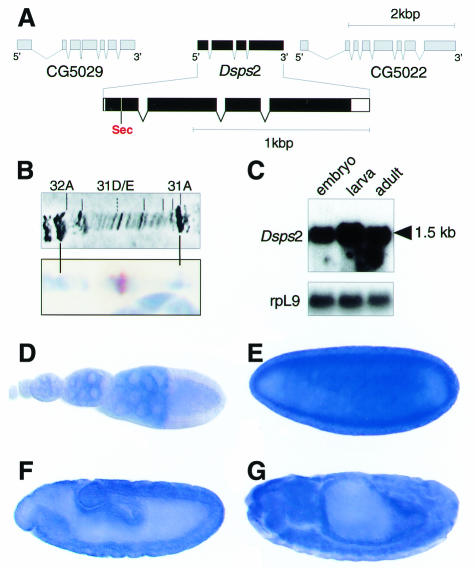
Fig. 1. Structure, location and expression of the Drosophila sps2 gene. (A) Genomic organization of Dsps2 showing that the gene is composed of four exons; coding sequences (black bars) and untranslated regions (open bars) are indicated. Note the position of the UGA in position 24 of the deduced protein. (B) In situ hybridization of Dsps2 cDNA to polytene chromosome showing a signal in section 31D/E (see text). (C) Northern blot showing a single transcript in poly(A)+ RNA of embryos, larvae and adults; rpL9 is a control for similar RNA loading. (D–G) In situ hybridization to ovaries (D), blastoderm (E), gastrula (F) and a germ band retracted embryo (G) showing that maternal Dsps2 transcripts are expressed in nurse cells (D) and that zygotic expression occurs in a spatially restricted pattern (see text). Orientation: anterior to the left, lateral view, dorsal is top (E–G). For staging see Campos-Ortega and Hartenstein (1985).

An official website of the United States government
Here's how you know
Official websites use .gov
A
.gov website belongs to an official
government organization in the United States.
Secure .gov websites use HTTPS
A lock (
) or https:// means you've safely
connected to the .gov website. Share sensitive
information only on official, secure websites.
