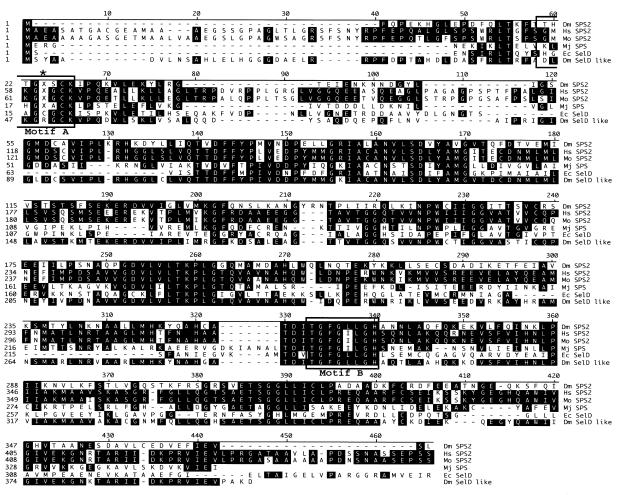
Fig. 2. Sequence comparison of DSPS2 and the known homologs. Alignment showing the Drosophila (Dm SPS2), human (Hs SPS2), mouse (Mo SPS2), M. jannaschii (Mj SPS), and E. coli (Ec SelD) SPS proteins and Drosophila SelD like protein (Dm SelD like). Position corresponding to Cys-17 of E. coli SelD is indicated by an asterisk, X denotes the Sec residue. Boxes: catalytic center (Motif A) and the ATP/GTP binding site (Motif B).

An official website of the United States government
Here's how you know
Official websites use .gov
A
.gov website belongs to an official
government organization in the United States.
Secure .gov websites use HTTPS
A lock (
) or https:// means you've safely
connected to the .gov website. Share sensitive
information only on official, secure websites.
