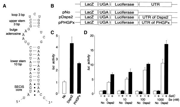
Fig. 3. 3′UTR-dependent readthrough of UGA in Drosophila tissue culture cells. (A) Predicted secondary structure of the 3′UTR of Dsps2 showing a form II SECIS element [see text and details in Grundner-Culemann et al. (1999)]. (B) Diagrams of transfection constructs for experiment. (C) Readthrough activity as determined for different reporter genes [for nomenclature see (B)] containing different 3′UTRs in transiently transfected Schneider cells. Luciferase activities were normalized versus β-galactosidase activity and ‘Luc activity’ is represented as a fold-increase over the baseline value which is given by No-UTR. (‘Luc activity’ mean values of six independent transfection experiments and standard deviation are indicated). Note higher Dsps2 3′UTR-dependent readthrough activity as compared to SECIS elements of mammalian glutathione peroxidase (pPHGPx). (D) Effect of selenium (adding sodium selenite to the medium; see Methods) or SelC (by overexpressing co-transfected DselC as indicated by a +; see Methods) on Dsps2 3′UTR-dependent readthrough activity.

An official website of the United States government
Here's how you know
Official websites use .gov
A
.gov website belongs to an official
government organization in the United States.
Secure .gov websites use HTTPS
A lock (
) or https:// means you've safely
connected to the .gov website. Share sensitive
information only on official, secure websites.
