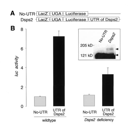
Fig. 4. SECIS element-dependent readthrough of the UGA stop codon in Drosophila embryos. (A) Schematic representation of the reporter genes used to construct transgenic embryos (see Methods). (B) Readthrough activity as determined from extracts of wildtype (left side) or Dsps2 deficiency mutant embryos (right side; see text; readthrough activity was determined as described in the legend of Figure 3). Relative activities represent mean values from six independent collections of embryos; standard deviations are indicated. Inset: western blot indicating that embryonic extracts contain a 3′UTR-dependent β-galactosidase/luciferase fusion protein as revealed by anti-β-galactosidase-antibody staining. The fusion protein and UGA-terminated β-galactosidase are indicated by arrowheads.

An official website of the United States government
Here's how you know
Official websites use .gov
A
.gov website belongs to an official
government organization in the United States.
Secure .gov websites use HTTPS
A lock (
) or https:// means you've safely
connected to the .gov website. Share sensitive
information only on official, secure websites.
