
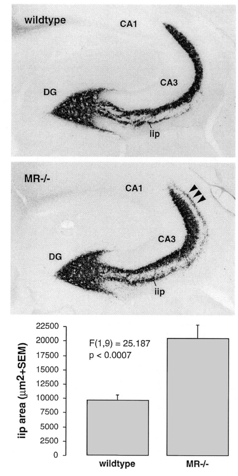
Fig. 2. Mossy fiber sprouting in the hippocampus of adult MR–/– mice. In comparison to wild-type mice with regular distribution of Timm-stained mossy fiber terminals, MR–/– mice demonstrate significantly prolonged intra/infrapyramidal projection fields (iip) that extend up to the distal end of CA3 (arrowheads). Quantitative morphometrical analysis confirmed that the intra/infrapyramidal projection field was on average twice as large in MR–/– mice, whereas the suprapyramidal and hilar projection fields were not significantly enlarged.