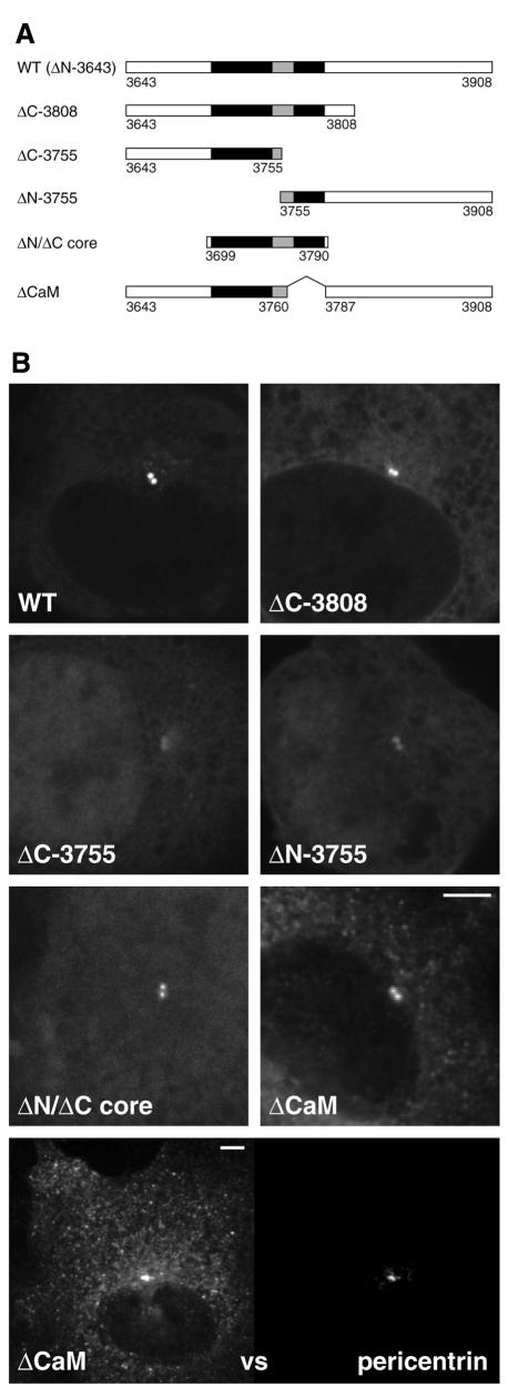
Fig. 3. At least two parts of the conserved region of the AKAP450 C-terminus contribute to centrosomal targeting. (A) The parts of the C-terminal region of AKAP450 fused to GFP in the indicated constructs. The residue numbers at the beginning and end of each construct are shown, with residue 3908 being the C-terminus of the whole protein. The two well conserved sections underlined in Figure 1B are shown in black. (B) COS cells expressing the six GFP fusions shown in (A). Cells were photographed using identical settings, and the perinuclear spots colocalized with endogenous pericentrin (not shown). In addition the lowest panel shows double labelling of endogenous pericentrin and ΔCaM at high levels of expression, the latter showing centrosomal staining and also a punctate pattern in the cytosol. Scale bars, 5 µm.

An official website of the United States government
Here's how you know
Official websites use .gov
A
.gov website belongs to an official
government organization in the United States.
Secure .gov websites use HTTPS
A lock (
) or https:// means you've safely
connected to the .gov website. Share sensitive
information only on official, secure websites.
