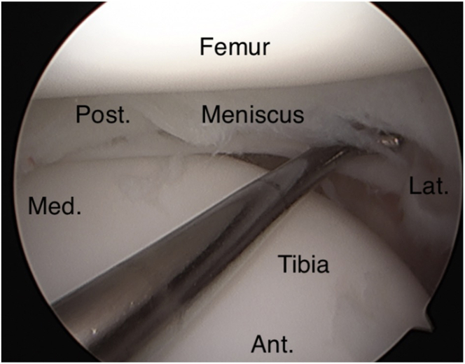
Fig 28.
Shown is the probe inspecting the lateral joint space including the lateral meniscus as a whole including the meniscus root, the lateral femoral chondral surface, the lateral tibial plateau chondral surface, and the popliteal tendon. In this case, a discoid lateral meniscus with anterior rim instability and tearing was identified. (Ant., anterior; Lat., lateral; Med., medial; Post., posterior.)

