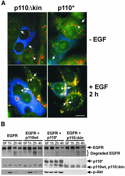
Fig. 1. EGFR endocytosis after inhibition of EGF-stimulated PI3K activity. (A) SKBR-3 cells were transfected with myc-tagged mutant p110Δkin or p110* for 48 h and then stimulated with EGF for 2 h or not stimulated. Indirect immunofluorescence was performed as described in Methods. Photographs are triple exposures with EGFR localization in the green channel (arrows), Lamp1 localization in the red channel (arrowheads) and transfected p110 in the blue channel. Size bar = 15 µm. (B) 293T cells were transfected with human EGFR, wild-type PI3K p110, mutant p110Δkin and/or p110* for 48 h and then stimulated with EGF for the indicated time. Cell lysates were subjected to immunoblot analysis with anti-EGFR, anti-myc or anti-phospho-Akt (p-Akt) antibodies.

An official website of the United States government
Here's how you know
Official websites use .gov
A
.gov website belongs to an official
government organization in the United States.
Secure .gov websites use HTTPS
A lock (
) or https:// means you've safely
connected to the .gov website. Share sensitive
information only on official, secure websites.
