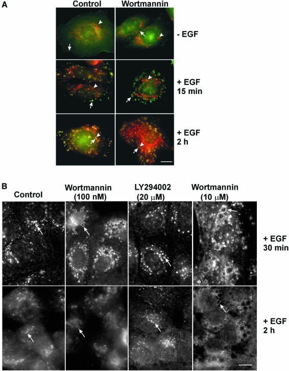
Fig. 2. Effects of wortmannin on the intracellular trafficking of EGFR in SKBR-3 and MDCK cells. (A) SKBR-3 cells. Left panel, control. Right panel, cells treated with wortmannin (100 nM). Cells were either kept in serum-deprived media or stimulated with EGF for 15 min or 2 h. Photographs are double exposures with EGFR localization in the green channel (arrows), Lamp1 localization in the red channel (arrowheads). (B) MDCK cells. Left most panels: control. Second panels: cells treated with wortmannin (100 nM). Third panels: cells treated with LY294002 (20 µM). Right most panels: cells treated with a high concentration of wortmannin (10 µM). Cells were stimulated with EGF for 30 min or 2 h. EGFR localization (arrows) was revealed by indirect immunofluorescence. Size bar = 20 µm.

An official website of the United States government
Here's how you know
Official websites use .gov
A
.gov website belongs to an official
government organization in the United States.
Secure .gov websites use HTTPS
A lock (
) or https:// means you've safely
connected to the .gov website. Share sensitive
information only on official, secure websites.
