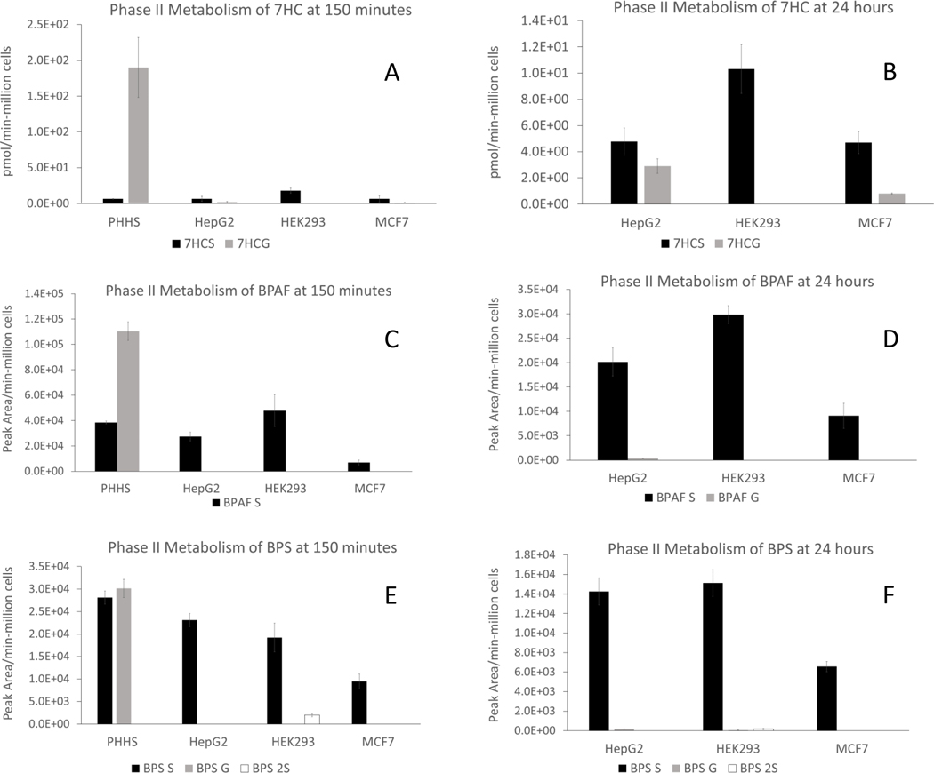
Figure 2. Glucuronide and sulfate conjugates assay in PHH and three Tox21 cell lines.
HepG2, HEK293 and MCF7 cells were incubated at 50,000 cells/well for 24 hours individually in comparison to PHHs (50,000 cells/well) incubated with 50 μM 7HC (A, 150 minutes; B, 24 hours); 50 μM BPAF (C, 150 minutes; D, 24 hours), or 50 μM BPS (E, 150 minutes; F, 24 hours). LC-MS assays were performed to quantify 7HC-glucuronide and sulfate conjugates, semi-quantify BPAF-glucuronide, sulfate conjugates, BPS-glucuronide and sulfate conjugates. Metabolite concentrations were converted to pmol of total metabolite per million cells plated for 7HC conjugates (pmol/min-106 cells) and peak area of total metabolite per million cells plated (peak area/min-106 cells) for BPAF and BPS conjugates.

