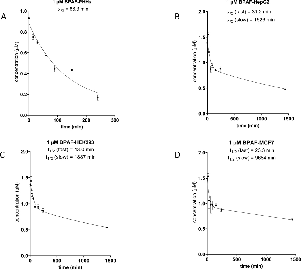
Figure 4. Clearance of BPAF by PHH and three Tox21 cell lines.
HepG2, HEK293 and MCF7 cells were incubated at 50,000 cells/well for 24 hours individually in comparison to PHHs (50,000 cells/well) incubated with 1.0 μM BPAF at 0, 15, 30, 60, 90, 150 and 240 minutes. LC-MS analysis was performed to monitor the clearance of BPAF parent compound at 1.0 μM to derive apparent half-life (t1/2). BPAF clearance in PHHs (A); BPAF clearance in HepG2 cells (B); BPAF clearance in HEK293 cells (C); BPAF clearance in MCF7 cells (D).

