Figure 1.
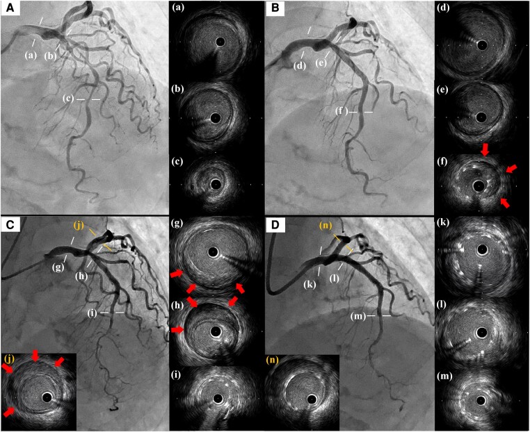
(A) Initial left coronary angiography from a straight cranial view and images of IVUS (a–c). IVUS demonstrated no haematoma but low-echoic plaque. (B) Post-primary PCI. IVUS revealed haematoma in the stent distal (red arrows) but not in the proximal vessel (d–f). (C) Emergent coronary angiography and IVUS on day seven. The previous haematoma extended to the LMT through the LAD (g–i) and even to the proximal LCX (j). Red arrows indicated the newly observed haematoma. (D) Final angiography and IVUS after the bailout PCI. Additional stents were implanted, covering the LMT to the LAD (k–m) and the LCX (n) using the T-stent methods. IVUS, intravascular ultrasound study; PCI, percutaneous coronary intervention; LMT, left main trunk; LAD, left anterior descending artery; LCX, left circumflex artery.

