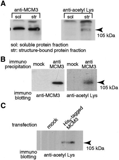
Fig. 1. In vivo acetylation of MCM3. (A) The soluble protein fraction and the structure-bound protein fraction prepared from 5 × 10⁵ asynchronous cells were analyzed by 7.5% SDS–PAGE. The proteins were blotted to PVDF membrane. The membranes were incubated with indicated antibody, respectively. The bound antibody was detected by horseradish peroxidase-labeled secondary antibody and visualized by ECL PLUS kit. (B) HeLa cell extracts prepared from 1 × 10⁷ cells were incubated with protein A–Sepharose and with anti-MCM3 antibody or normal rabbit IgG (mock). After 1 h incubation, column-bound proteins were analyzed by SDS–PAGE (7.5%) and blotted to PVDF membrane. (C) MCM3/pcDNA3.1 plasmid or mock vector was transfected to 293T cells, respectively. After culture for 20 h the cells were harvested and resuspended in hypotonic buffer containing 1% NP40, 0.5 M NaCl, and 7 mM Na butyrate. Cell extracts were incubated with a TALON column and the column was washed extensively with same buffer. Bound proteins were extracted from the column with 100 mM imidazole and 7 mM Na butyrate in hypotonic buffer and analyzed by SDS–PAGE. The analyzed proteins were blotted to PVDF membrane.

An official website of the United States government
Here's how you know
Official websites use .gov
A
.gov website belongs to an official
government organization in the United States.
Secure .gov websites use HTTPS
A lock (
) or https:// means you've safely
connected to the .gov website. Share sensitive
information only on official, secure websites.
