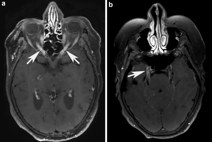
Fig. 3.
Magnetic resonance imaging findings of anti-GFAP meningoencephalomyelitis – autopsy case #2 Brain in vivo MRI from autopsy patient #2 shows a bilateral optic neuritis (a, white arrows) on post-contrast axial angulated reconstructions of T1-weighted blackblood-images. Further axial angulated reconstruction of post-contrast T1-weighted blackblood-images shows enhancement throughout the cisternal segment of both trigeminal nerves (b). The right trigeminal nerve additionally shows an abnormal enhancement in the trigeminal cave (b, white arrow), indicating an involvement of the ganglion

