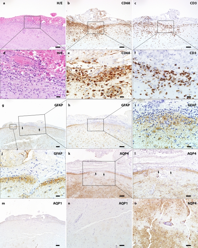
Fig. 8.
Subpial pathology and parenchymal aquaporin loss in GFAP autoimmunity (a) H&E staining shows hypercellularity in the leptomeninges and subpial parenchyma. Increased collagen is noted in the leptomeninges (magnified view in panel d). (b) CD68 on the consecutive section highlights marked macrophages/microglia in both meninges and subpial parenchyma (high power view in panel e). (c) Infiltration of T lymphocytes in meninges and towards the deep parenchyma is seen in the same region (magnified view in panel f). (g-j) GFAP staining indicates alteration of glia limitans in the inflamed area. (h) Decreased GFAP along pial surface. (i) The high-power view highlights discontinuous glia limitans and some adjacent hypertrophic astrocytes. (j) The adjacent non-inflamed region shows intact glia limitans. (k) Immunohistochemistry indicates decreased pial AQP4 immunoreactivity in the highly inflamed region (high power view in l). (m) Decreased pial AQP1 is also noted. (n) Cortical perivascular loss of AQP1 immunoreactivity. (o) Perivascular decreased AQP4 immunoreactivity. Scale bars a-c 100 µm; g, m 200 µm; l, n, o 50 µm; h, k 100 µm; d-f, i, j 20 µm. AQP1 aquaporin 1, AQP4 aquaporin 4, CD cluster of differentiation, GFAP glial fibrillary acidic protein, H&E hematoxylin & eosin

