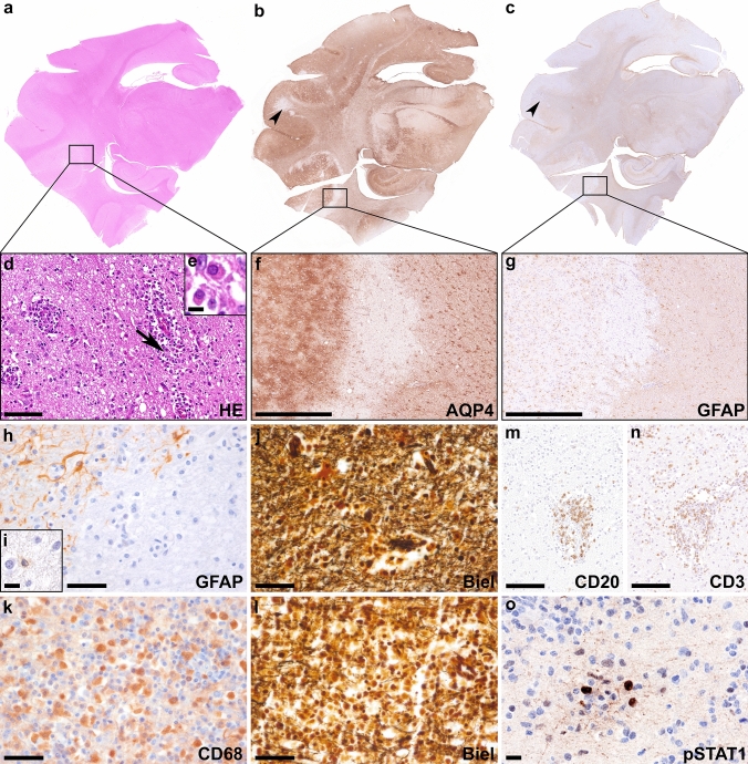
Fig. 9.
Neuropathological findings in a canine autopsy case with anti-GFAP autoantibodies Overview stainings (H&E (a); AQP4 (b); GFAP (c)) of the same area of a canine temporal brain section including the hippocampus. H&E staining (d) shows edema, reactive gliosis, neutrophilic granulocytes and mitoses and perivascular inflammatory infiltrates mainly consist of lymphocytes and plasma cells (arrow in d enlarged in inset e). In some parts of the lesions AQP4 (f) and GFAP (g, h) are selectively lost (h) with a few apoptotic astrocytes remaining within the lesion (i), while axons are relatively well preserved (j; Biel). Other parts of the lesions showed focal cystic necrosis with abundant CD68 positive macrophages (k) and loss of axons (l; Biel). Immunohistochemistry reveals some CD20+ B cells (m) around vessels. Inflammation is characterized by an abundant number of perivascular and parenchymal CD3+ T cells (n). pSTAT1 is mainly upregulated in lymphocytes (o). Scale bars d, m, n 50 µm; f, g 500 µm; e, i, o 10 µm; h, j-l 25 µm. AQP4 aquaporin 4, Biel Bielschowsky, CD cluster of differentiation, GFAP glial fibrillary acidic protein, H&E hematoxylin & eosin, pSTAT1 phosphorylated signal transducer and activator of transcription 1

