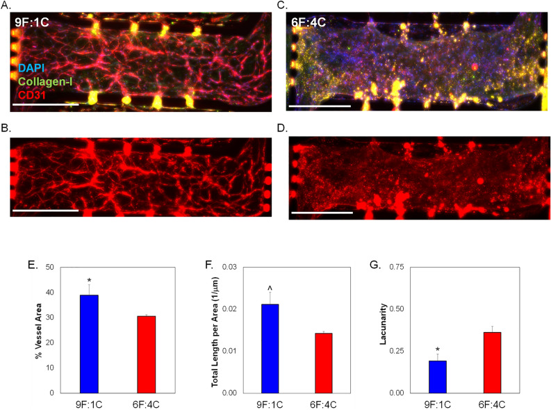
Figure 2.
Analysis of microvessel formation in microtissue models testing varying collagen I concentrations. Total ECM content was held constant at 10 mg/mL, while the ratio of fibrin to collagen - was varied. 9 parts fibrin to 1 part collagen is abbreviated 9F:1C; with 6F:4C representing 6 parts fibrin to 4 parts collagen-I. No cancer cells were loaded in side chambers for these experiments, to focus on vascularization potential only of the different matrix combinations. Representative images of center microtissues of devices with (A,B) 9F:1C and (C,D) 6F:4C. Immunostaining includes DAPI, CD31 (TXRED), and collagenI (GFP). Scale bars = 200 µm. (E) Quantification of average blood vessel area in center chambers. (F) Total length of vessels per area in side chambers. (G) Average lacunarity of vessels in microtissues. Data shown as average + SEM, N = 3. Samples compared via Mann–Whitney paired tests. *p < 0.05; ^ p = 0.06.

