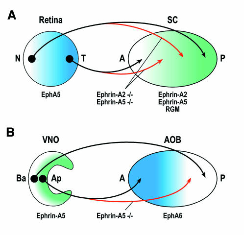
Fig. 1. Topographic mapping by Ephrin-A ligands and their EphA receptors in the mammalian visual system (A) and vomeronasal system (B). SC, superior colliculus; N, nasal; T, temporal; A, anterior; P, posterior; VNO, vomeronasal organ; AOB, accessory olfactory bulb; Ba, basal; Ap, apical. Black shows the wild type projection patterns, red shows typical projection errors in Ephrin-A2, Ephrin-A5 double mutant mice (A) or Ephrin-A5 single mutants (B).

An official website of the United States government
Here's how you know
Official websites use .gov
A
.gov website belongs to an official
government organization in the United States.
Secure .gov websites use HTTPS
A lock (
) or https:// means you've safely
connected to the .gov website. Share sensitive
information only on official, secure websites.
