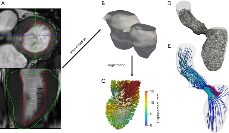
Figure 3.
General image-based computational procedure for patient-specific simulations. From diagnostic imaging data (A), segmentation yields anatomical surfaces (B) possibly for different times of the heartbeat. Registration algorithms are used to reconstruct displacement fields (C) with respect to a reference configuration. A computational mesh of the patient’s anatomy is generated (D) and used to perform numerical simulations (streamlines of blood flow in left ventricle and aorta) (E). Credits: M. Fedele, I. Fumagalli.

