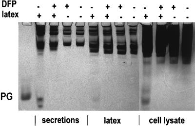
FIG. 5.
AU-PAGE of neutrophil secretions stimulated by latex beads. Gels were stained with Coomassie blue. Each lane contains either 107 cell equivalents of either neutrophil secretions, extracellular latex beads after phagocytosis, or neutrophil lysates. The symbols + and − indicate which materials were added into each tube before the phagocytosis started. At the end of the incubation, DFP and/or latex beads were added into tubes that did not have them during phagocytosis. The PG standard is 0.5 μg of PG3.

