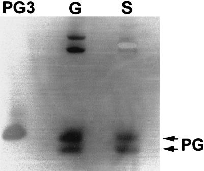
FIG. 6.
Western blot analysis of neutrophil secretions stimulated by latex beads. After AU-PAGE, proteins were blotted to an Immobilon-P membrane, and the membrane was probed with a monoclonal anti-PG3 antibody and rabbit anti-mouse immunoglobulin G-alkaline phosphatase conjugate, then developed in 5-bromo-4-chloro-3-indolylphosphate–nitroblue tetrazolium solution. PG3, 0.5 μg of synthetic PG3; G, porcine neutrophil granule lysate (5 × 10⁶ cell equivalents); S, neutrophil secretions generated during phagocytosis of latex beads in the absence of DFP (107 cell equivalents).

