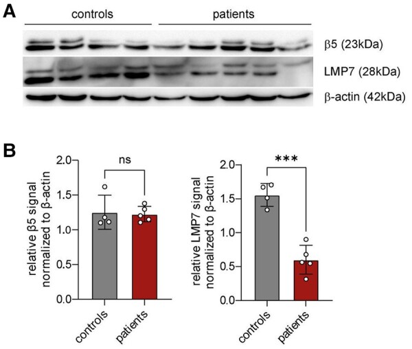
Figure 3.

An analysis of immunoproteasome expression in patients with TLE. (A) A western blot analysis of immunoproteasome subunit LMP7 and constitutive proteasome subunit β5 in female patients with epilepsy (n = 5) compared with controls (n = 4). (B) The bar graphs show relative β5 and LMP7 signals normalized to β-actin. The statistical analysis was performed by using an unpaired t-test (n.s. = not significant; ***P < 0.001). Data are presented as mean ± SD. Uncropped blots are shown in Supplementary Fig. 6.
