
Figure 2. Proliferative capacity of antigen specific T cells.
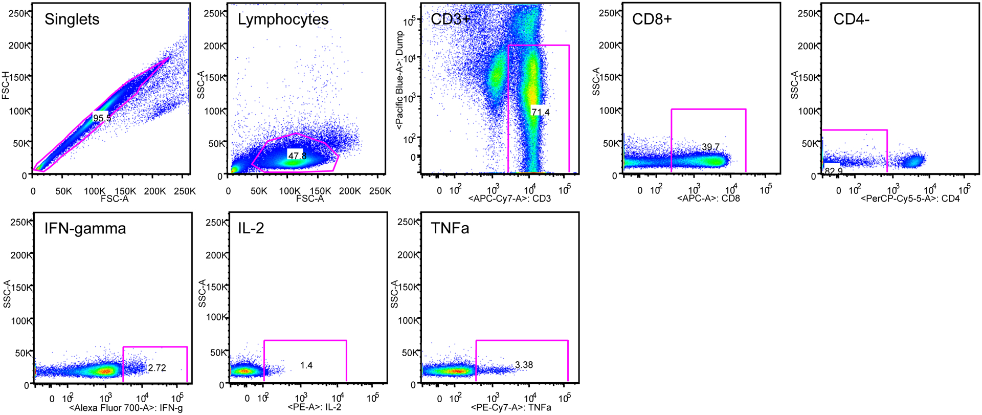
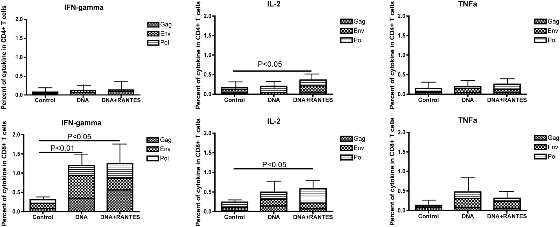
PBMCs from immunized macaques are stained with CFSE and stimulated with growth medium, concanavalin A (ConA), or SIV gag, SIV pol, or HIV Env peptides for 5 days. Following stimulation, cells were stained for phenotypic markers and analyzed by flow cytometry. A) Average proliferative capacity of CD4 and CD8 T cells B) Average for all primates is presented for CD4 or CD8 TCMs and TEMs. Data were acquired on an LSRI instrument analyzed with FlowJo software.