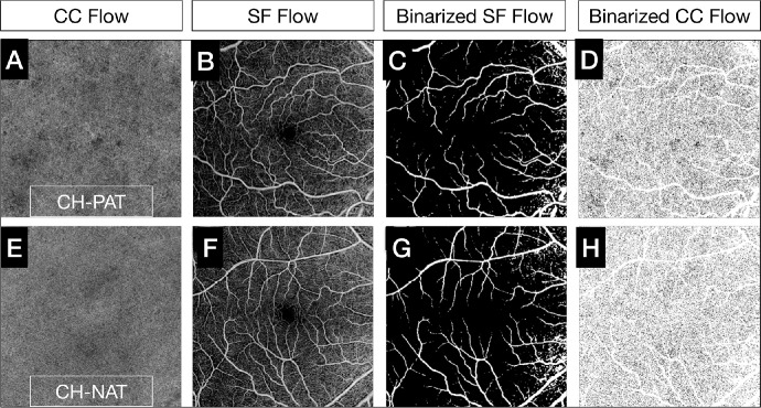
Figure 4.
Comparison of CC flow deficits between CH-PAT and CH-NAT subjects. Customized CC en face OCTA slabs segmented just below BM (4–20 µm) were generated (A, E). The default superficial en face OCTA slabs (B, F) were binarized (C, G) and overlapped to the binarized CC en face OCTA slab to mask the projections of superficial and large vessels, which were excluded from the analyses. The final binarized CC en face OCTA slab used to compute the CC FD% is illustrated in D and H, and the CC en face OCTA slab from a CH-PAT eye shows increased CC FDs (D) compared to a CH-NAT eye (H).

