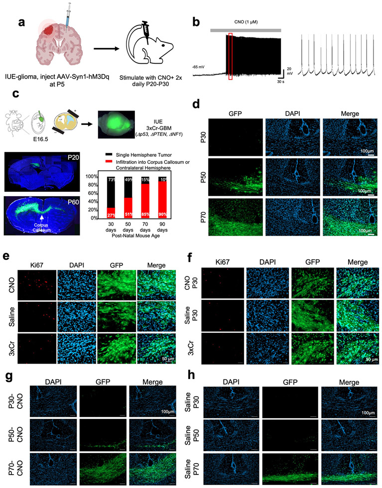
Extended Data Figure 2. Contralateral cortical stimulation accelerates tumor progression.
a. Schematic depicting injection of AAV into the contralateral cortex. b. Electrophysiology measuring neural activity in response to CNO treatment on mouse brain sections to confirm DREADD activity. c. Schematic of intra-uterine electroporation (IUE) model of high-grade glioma (HGG). Representative images of tumor brains from 20 day old mice versus 60 days old. Tumor brain slices were stained with Hoechst, and native GFP fluorescence was utilized to visualize tumor. Time course of contralateral cortical infiltration. Log-regression of 3xCr tumors demonstrated that infiltration correlates strongly with time (two-sided log-regression, Chi-square = 23.38, df = 1, p-value <0.0001, n30 =22, n50=19, n70=21, n90=10). 72 IUE-HGG bearing mice were generated from 10 separate litters of mice and sampled across P30, P50, P70, P90 to ensure accurate representation of infiltration. d. Representative images of infiltrating tumor across the midline at each respective timepoint. e. Representative Ki67 staining of CNO-treated versus Saline-treated versus untreated IUE-HGG sections taken from P30 tumor brains. f. Representative Ki67 staining of CNO-only control, without hM3Dq, Saline, and IUE-HGG sections taken from P30 tumor brains, g-h. Representative images of infiltrating tumor across the midline at each respective timepoint from the CNO-only control experiments (g) and saline only control (h). Tumor sections were imaged and quantified for each condition from at least five mice to ensure reproducibility (d-h).

