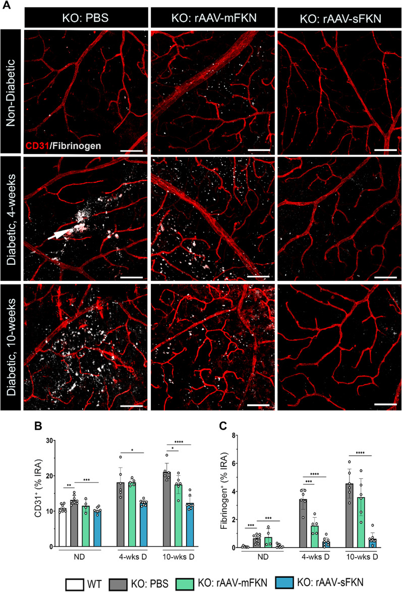
Fig. 3.
Intra-vitreal administration of rAAV–sFKN reduces vascular pathology and fibrin(ogen) deposition in the diabetic retina. A Confocal images of retinal tissues stained for CD31 (red) and fibrinogen (white) (Scale bar; 100 µm). B, C Quantification of retinal immunofluorescence analysis of CD31+ percent immunoreactive area (% IRA) (B) and fibrinogen+ immunoreactive area (% IRA) (C). Data shown as mean ± SD, n = 4–8 mice per group, where each dot represents an individual mouse across six experiments. *p < 0.05, **p < 0.01, ***p < 0.001 and ****p < 0.0001 using Student’s t test, Welch’s correction

