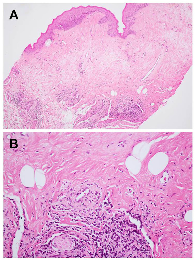
Figure 2.

Histopathological findings (hematoxylin-eosin): (A) focal atrophic epithelium with hydropic degeneration of the basal cell layer and edematous and homogenized collagen fiber in the superficial lamina propria (original magnification x100), (B) patchy, band-like lymphoplasmacytic inflammatory infiltration below the hyalinized area (original magnification x400).
