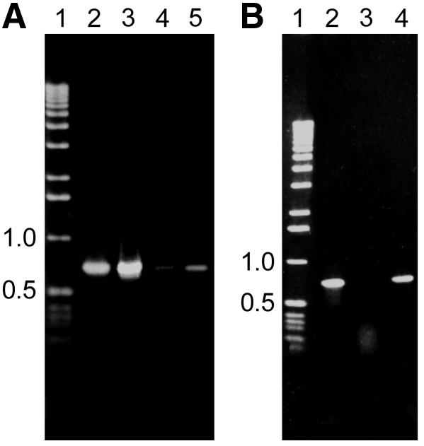
Fig. 2. PCR analysis from DNA bound to HAP or the supernatant after crosslinking of CHO cells transfected with either pEPI-1 or pGFP-C1. (A) PCR analysis of HAP-bound DNA. Lane 1, molecular weight marker; lane 2, control PCR using 1 ng of pEPI-1 vector DNA as a template; lane 3, PCR analysis from pEPI-1 transfected cells using 10 ng of DNA as a template; lane 4, PCR analysis from pGFP-C1 transfected cells using 10 ng of DNA as a template; lane 5, as lane 4 but using 100 ng of DNA as a template. (B) PCR analysis of the supernatant from pEPI-1 transfected cells using 10 ng of DNA as a template. Designations are as in (A).
