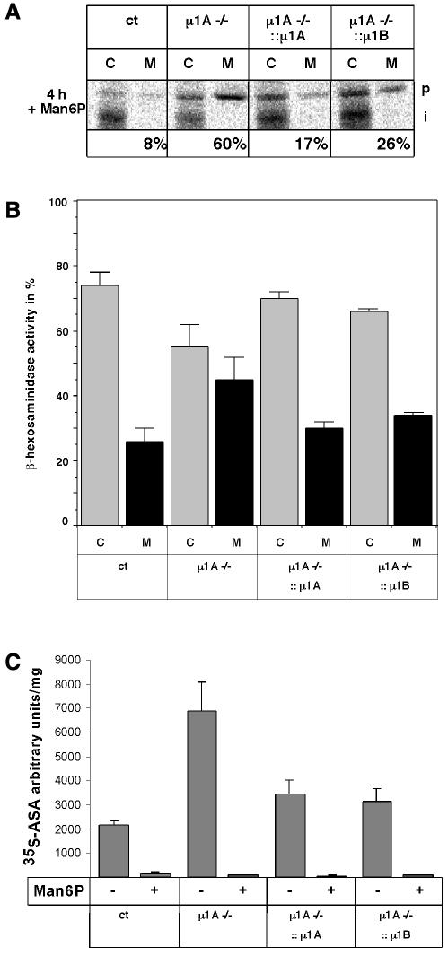
Fig. 5. Cathepsin D and β-hexosaminidase intracellular sorting in control (ct), µ1A–/–, µ1A–/–::µ1A and µ1A–/–::µ1B cells. (A) Man6P was added to the medium to prevent endocytosis of secreted cathepsin D. Metabolically labelled cathepsin D was immunoprecipitated from cell extracts (C) and from the medium (M). The Golgi precursor form (p) and the endosomal intermediate processed form (i) are indicated. (B) Secretion of β-hexosaminidase to the medium. Shown here is the distribution of the enzymatic activity between the cells and the medium as a percentage of the total (n = 2). (C) Endocytosis of arylsulfatase A (ASA) by control (ct), µ1A–/–, µ1A–/–::µ1A and µ1A–/–::µ1B cells (n = 2). Arbitrary units correspond to pixel numbers on phosphoimager screens (Fuji BAS1000).

An official website of the United States government
Here's how you know
Official websites use .gov
A
.gov website belongs to an official
government organization in the United States.
Secure .gov websites use HTTPS
A lock (
) or https:// means you've safely
connected to the .gov website. Share sensitive
information only on official, secure websites.
