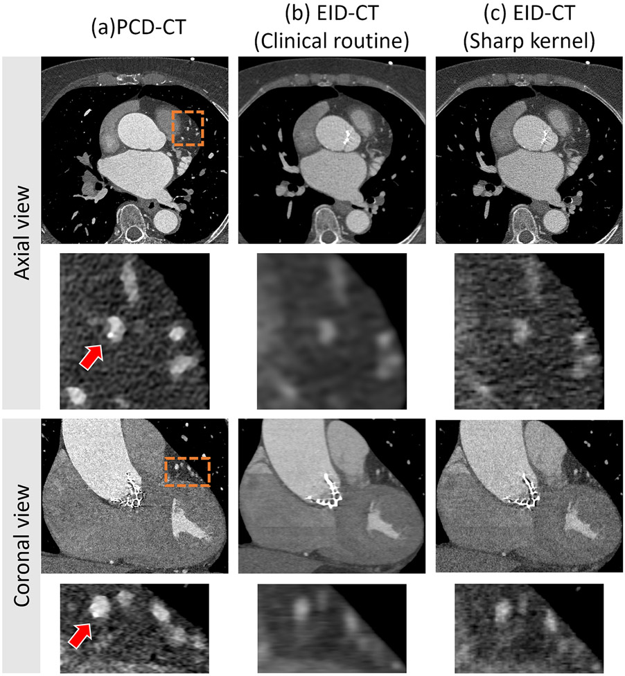
Figure 4.
Sample images of small calcium detection in patients. Axial and coronal view of PCD UHR (a), EID-clinical routine (b), and EID-sharp kernel (c) CT images of a coronary CTA in a 77-year-old man show that high spatial resolution enabled detection of the very small calcification (arrow in (a)). Image display window (WL/WW: 200/1000 HU).

