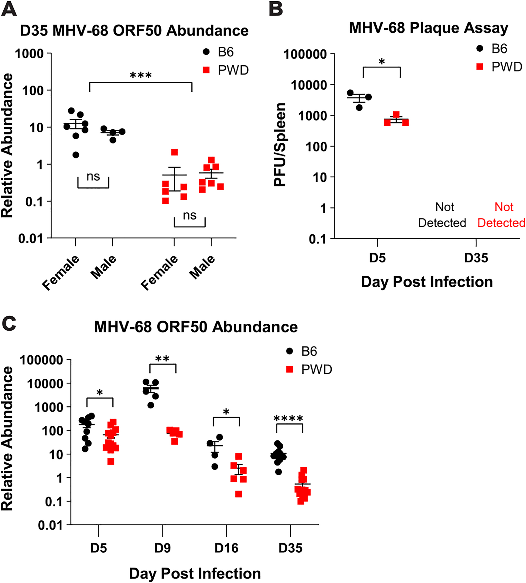
Figure 5. PWD mice demonstrate lower viral load across MHV-68 infection.

Female and male 6–8 week old B6 and PWD mice were i.p. infected with MHV-68 as described in Figure 1. At 5- (D5) (9F B6 and 6F and 7M PWD, pooled across two experiments), 9- (D9) (3F + 2M B6 and 3F + 2M PWD), 16- (D16) (2F + 2M B6 and 4F + 2M PWD), or 35- (D35) (7F + 4M B6 and 6F + 7M PWD pooled across two experiments) days post infection splenic tissue was harvested and processed for DNA isolation and quantification of viral load by MHV-68 ORF50 qPCR, or determination of infectious viral titers by plaque assay (see Materials and Methods). (A) MHV-68 ORF50 relative abundance (normalized by host DNA abundance) in B6 and PWD mice at the timepoint of transcriptome analysis (D35). Significance of differences between B6 and PWD genotypes, as well as differences between males and females within genotype, were determined by two-way ANOVA with Šídák’s multiple comparisons test. (B) Comparison of plaque forming units (PFU) per spleen in infected B6 and PWD mice, sexes pooled, at D5 and D35. (C) Analysis of viral load in B6 and PWD mice, sexes pooled, across the course of MHV-68 infection via MHV-68 ORF50 DNA relative abundance. Significance of differences between B6 and PWD genotypes at each infection time point were determined by multiple unpaired T-tests with a P value threshold of 0.05. Comparisons are indicated by the brackets where significant (p<0.05).
