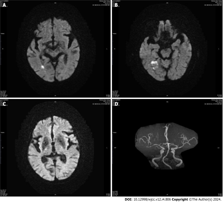
Figure 1.
Neuroimaging of Patient 1's first hospitalization. A-C: Diffusion-weighted imaging showing restricted diffusion in the right temporal lobe, and the basal ganglia, thalamus, and subthalamic nucleus were entirely spared; D: Magnetic resonance angiography of the head showed significant stenosis of the right middle cerebral artery (MCA) and mild stenosis of the left MCA.

