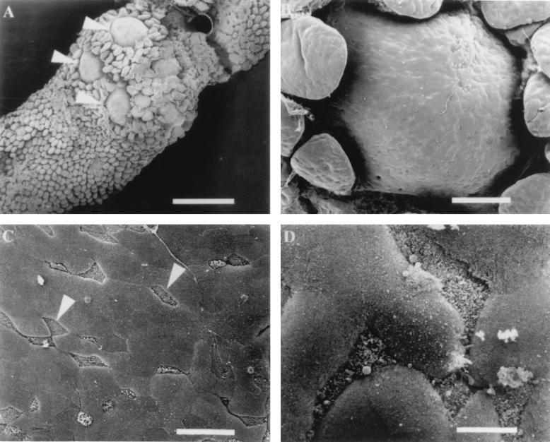
FIG. 1.
Scanning electron micrographs of murine Peyer’s patch tissue. (A) Luminal side of Peyer’s patch containing several domes (arrowheads) surrounded by villi. Bar, 700 μm. (B) Epithelial surface of a lymphoid follicle dome that contains numerous M cells and enterocytes randomly distributed. Bar, 100 μm. (C) Region of the FAE containing several M cells (arrowheads) that can be distinguished by short microvilli which give an irregular appearance. Bar, 20 μm. (D) Higher magnification of M cells and enterocytes. M cell microvilli create a rough irregular appearance in comparison to adjacent enterocytes which look smooth in texture. Bar, 6 μm.

