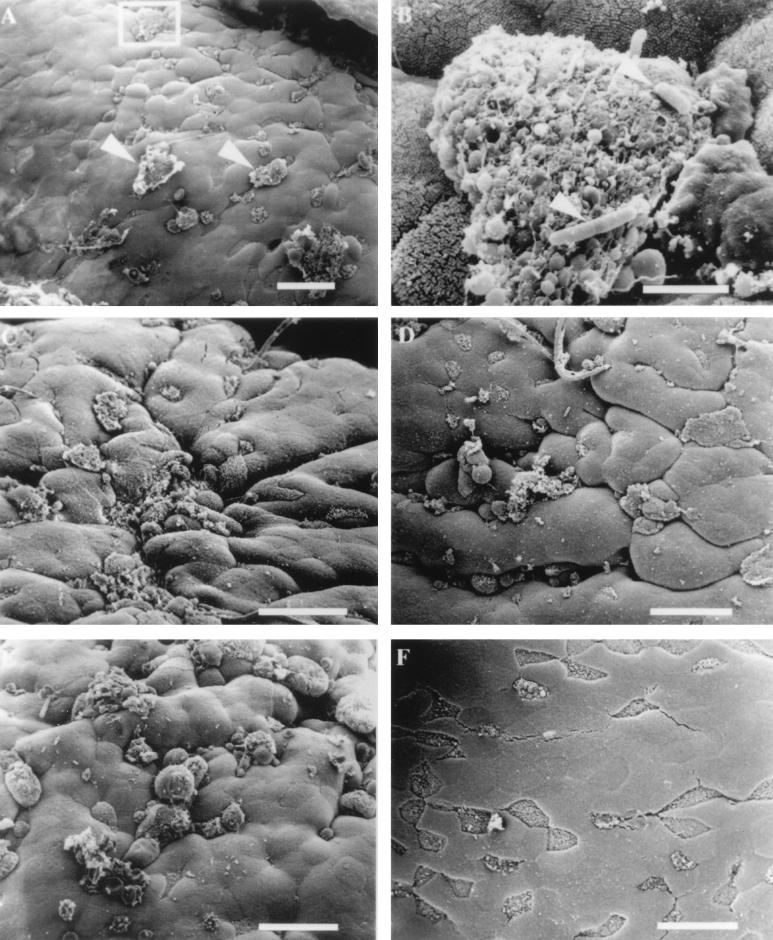
FIG. 2.
Electron micrographs of mouse FAE infected with invasive S. typhimurium SL1344. (A) Membrane rearrangements are visible on the surfaces of some M cells (arrowheads) 30 min after infection with S. typhimurium SL1344. (B) Higher magnification of the region in the inset from panel A. Rod-shaped organisms (arrowheads) can be observed adhering to M cell debris. Bar, 3 μm. (C) Effects of S. typhimurium 60 min after infection. (D) Tissue infected for 90 min with invasive Salmonella. M cells with a normal apical appearance are difficult to find, and the majority of cells display membrane disruptions and blebbing. (E) Infection of ligated loop with invasive Salmonella for 120 min. A quantitative increase in the number of M cells displaying membrane changes and a qualitative increase in the size of apical membrane disruption are evident. (F) Mouse intestinal tissue inoculated with bacterial growth broth for 30 to 120 min was examined for the appearance of uninfected tissue. The tissue shown has been inoculated with broth for 120 min and appears completely normal. The M cells are clearly visible, and the microvilli have the typical appearance of uninfected tissue. Bar, 20 μm unless otherwise indicated.

