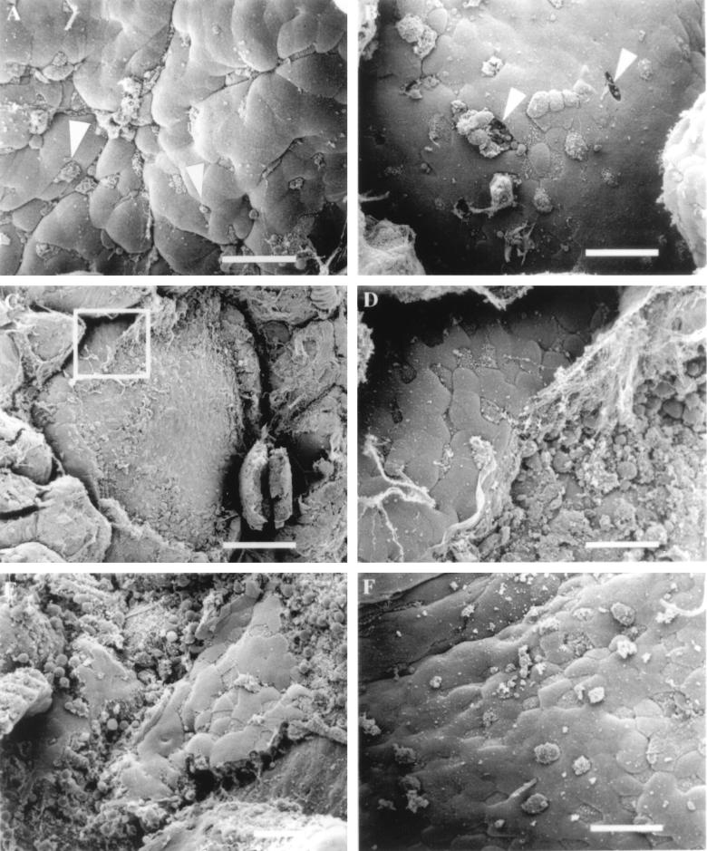
FIG. 3.
Effect of L. monocytogenes on murine FAE. (A) Mouse tissue infected for 30 min with L. monocytogenes 10403S. Some M cells display aberrant microvilli that are rounded in appearance (arrowheads). (B) After 60 min of infection, the presence of the bacteria appears to have destroyed the integrity of the FAE by creating holes (arrowheads) in the dome epithelium where M cells once resided. (C) Listeria-induced destruction of the FAE 90 min postinfection. Bar, 100 μm. (D) Higher-magnification view of the inset in Fig. C. At the periphery of the dome, a few M cells and enterocytes are still intact. (E) Tissue infected for 120 min with L. monocytogenes. (F) Tissue infected with the L. monocytogenes listeriolysin O-negative mutant DP-L2161 did not induce epithelial necrosis after 120 min of infection. Bar, 20 μm (unless otherwise indicated).

