
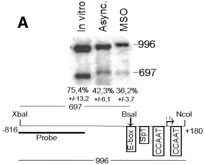
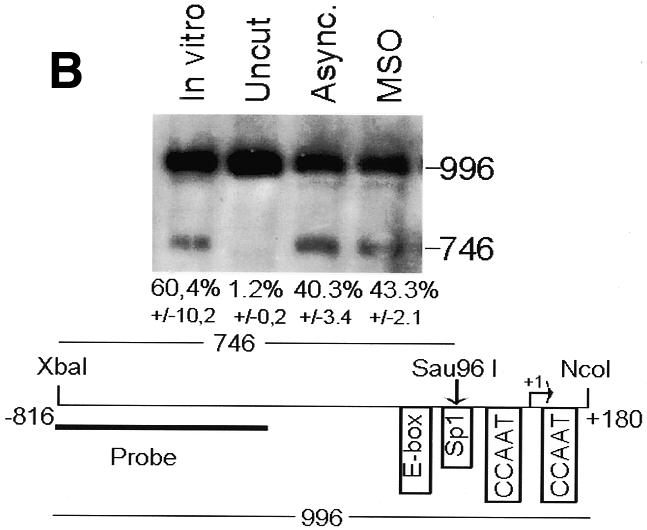
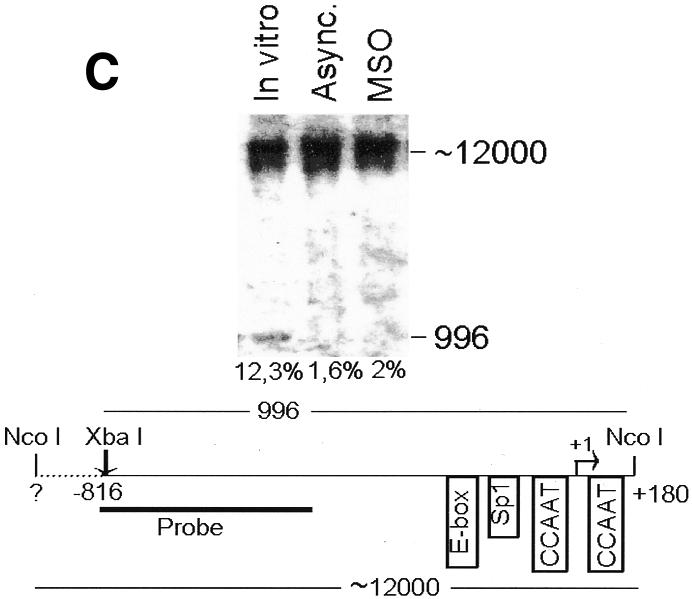
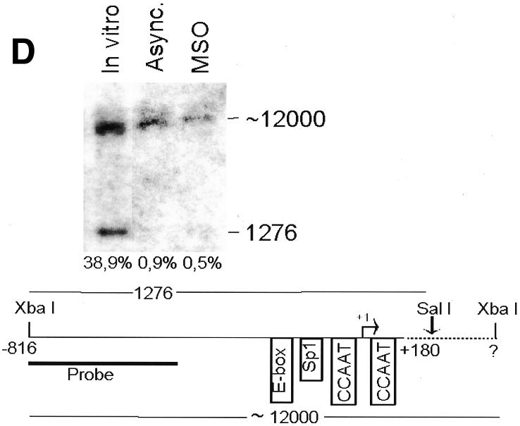
Fig. 2. The cyclin B1 promoter is accessible to restriction endonucleases in vivo. Nuclei isolated from asynchronous (Async.) (20 µg of DNA) and permeabilized MSO cells were partially digested with 100 U of BsaAI (A), 100 U of Sau96I (B), 200 U of XbaI (C) or 100 U of SalI (D). Purified DNA was fully digested with NcoI and XbaI when BsaAI was used for the partial digestion, and with NcoI when XbaI was used for the partial digestion. The probe used for the hybridization is shown at the bottom of each panel. Arrows mark the digested and undigested bands. The schematic diagram at the bottom of each panel indicates the position of the digested bands in the cyclin B1 promoter. The percent digestion is shown below each lane.