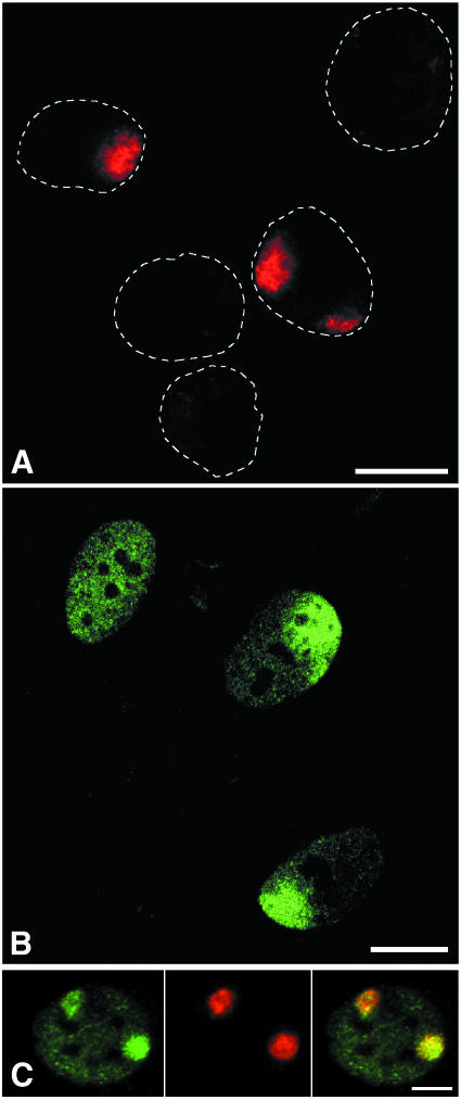
Fig. 1. (A) Detection of locally induced UV damage in cell nuclei. A UV-blocking polycarbonate filter containing pores of 5 µm in diameter was used to cover a monolayer of cells. The filter-covered cells were UV irradiated with 30 J/m2 and CPDs were subsequently detected by immunofluorescent labelling. Dotted lines denote the contours of individual cell nuclei. Two nuclei show labelling of CPDs in discrete nuclear areas. (B) Effect of local nuclear UV damage on the distribution of TFIIH. Human primary fibroblasts were locally UV irradiated with 100 J/m2 UV light, using a filter with 10 µm pores. Following irradiation, cells were grown for 1 h and immunolabelled against the p62 subunit of TFIIH. The top-left nucleus displays the characteristic labelling pattern of TFIIH in unirradiated cells, whereas the two remaining nuclei exhibit a TFIIH accumulation in UV-damaged nuclear areas, and a reduction in TFIIH signal outside these areas. A single confocal optical section is shown. (C) Colocalization of TFIIH (green) and CPDs (red). Human primary fibroblasts were locally irradiated with 30 J/m2 UV light, using a filter with 8 µm pores. Following irradiation, cells were grown for 30 min and dual labelled against both CPDs and the p62 subunit of TFIIH. Bars represent 10 µm.

An official website of the United States government
Here's how you know
Official websites use .gov
A
.gov website belongs to an official
government organization in the United States.
Secure .gov websites use HTTPS
A lock (
) or https:// means you've safely
connected to the .gov website. Share sensitive
information only on official, secure websites.
