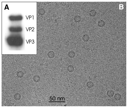
Fig. 1. (A) Western blot analysis of empty capsids. VP1, VP2 and VP3 were detected using a monoclonal antibody that recognizes all three capsid proteins. (B) Micrograph of unstained AAV2. The micrograph was recorded on a Philips CM 120 Biotwin at a nominal magnification of 52 000 with a defocus of 1900 nm. Particles appear as darker rings.

An official website of the United States government
Here's how you know
Official websites use .gov
A
.gov website belongs to an official
government organization in the United States.
Secure .gov websites use HTTPS
A lock (
) or https:// means you've safely
connected to the .gov website. Share sensitive
information only on official, secure websites.
