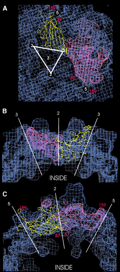
Fig. 4. Superposition of the Cα-chain (yellow) of the canine parvovirus VP2 capsid protein (PDB entry 1C8D) and the calculated surface of the VP2 capsid protein (pink) to the three-dimensional map of AAV2 (blue). The position of some residues of CPV-VP2 are labelled in red. Part of the inner surface of the capsid is shown in (A) with an almost straight on view to the eight-stranded β-barrel motif of CPV-VP2. The approximate positions of the symmetry axes are indicated in white. The views in (B) and (C) show different cross section through the protein shell.

An official website of the United States government
Here's how you know
Official websites use .gov
A
.gov website belongs to an official
government organization in the United States.
Secure .gov websites use HTTPS
A lock (
) or https:// means you've safely
connected to the .gov website. Share sensitive
information only on official, secure websites.
