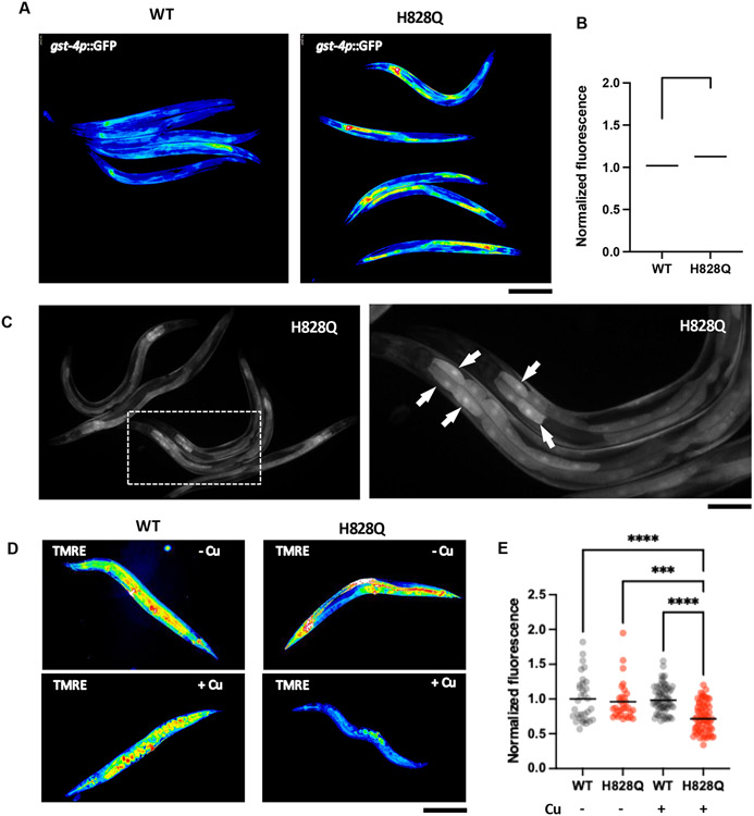
Figure. 3. Copper induces oxidative stress and mitochondrial damage in cua-1(knu790[H828Q]) strain.
A-C. Wild type (WT) and mutant (H828Q) animals were crossed with C. elegans strain expressing oxidative s tress sensor gst-4p::GFP. Resulting strains were synchronized, grown for 48h and incubated with CuCl2 overnight. Then gst-4p::GFP signal was analyzed by fluorescent microscopy. False color images (A) and quantification (B) show elevated gst-4p::GFP fluorescence in mutants compared to WT animals (** p<0.01, t-test; n≥60 animals) indicating higher oxidative stress in mutant s train. Panel C show distribution of gst-4p::GFP across the animal body in cua-1(knu790[H828Q]) strain. Right image shows higher magnifications of the boxed area in the left image. Arrows indicate elevated gst-4p::GFP signal in the cells laying in the proximal portion of the gut. D, E. WT and H828Q animals were grown in control or CuCl2-supplemented plates for 3 days and then stained with mitochondrial transmembrane potential sensor TMRE, whose intensity was analyzed by fluorescent microscopy. False color images of TMRE signal (D) and quantification (C) show reduced TMRE fluorescence in Cu-treated mutants compared to WT animals (**** p<0.0001, *** p<0.001, two way ANOVA; n≥30 animals) indicating a higher degree of mitochondrial damage in mutants train upon exposure to Cu. Scale bars: 300 μM (A; C, left panel; D), 160 μM (C, right panel).

