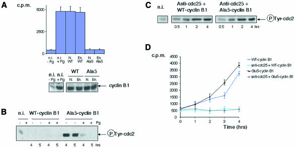
Fig. 1. Cyclin B1 phosphorylation is required for MPF activation in Xenopus oocytes. (A) Intact nucleated oocytes (N.) or enucleated oocytes (En.) were microinjected with either WT-B1 (WT) or Ala5-B1 (Ala5) mRNA (constructs containing a C-terminal VSVG-tag). Five hours later, groups of three oocytes were collected, lysed and immunoprecipitated with anti-P5D4 monoclonal antibodies. The beads were then analysed by H1 kinase assay (upper panel) or western blotting for cyclin B1 content (lower panel). Non-injected oocytes (n.i.), stimulated (+Pg) or not (–Pg) with progesterone, were used directly for H1 kinase assay or western blotting. The equivalent of one oocyte was used for each assay. Relative levels of cyclin B1 and B2 were shown recently to be a variable feature of different batches of mature oocytes, cyclin B1 often exceeding cyclin B2 by far (see figure 6 in Hochegger et al., 2001). Assuming that cyclin-B1–cdc2 and cyclin-B2–cdc2 complexes contribute to equivalent levels to H1 kinase activity in such oocytes, as reported in an earlier study (Kobayashi et al., 1991), exogenous cyclin B1 should not have been expressed to levels exceeding twice the endogenous cyclin B1 level in this experiment. (B) Oocytes were microinjected as in (A) (with higher levels of cyclin B1 expression) and treated or not with progesterone. At the indicated times, groups of three oocytes were homogenized and submitted to an immunoprecipitation with anti-P5D4 antibodies, followed by a western blot for the tyrosine-phosphorylated form of cdc2. Non-injected oocytes were used directly for western blotting. The equivalent of one oocyte was loaded per lane. (C) Oocytes were microinjected with affinity-purified anti-cdc25 antibodies and either WT-B1 or Ala5-B1 mRNA. Groups of three oocytes were lysed and analysed by western blotting for tyrosine-phosphorylated cdc2 content. The equivalent of one oocyte was loaded per lane. (D) Oocytes were microinjected or not with anti-cdc25 antibodies and with either WT-B1 or Glu5-B1 mRNA. Extracts from individual oocytes selected at the indicated times after microinjection were assayed for H1 kinase. The error bars represent standard deviation.

An official website of the United States government
Here's how you know
Official websites use .gov
A
.gov website belongs to an official
government organization in the United States.
Secure .gov websites use HTTPS
A lock (
) or https:// means you've safely
connected to the .gov website. Share sensitive
information only on official, secure websites.
