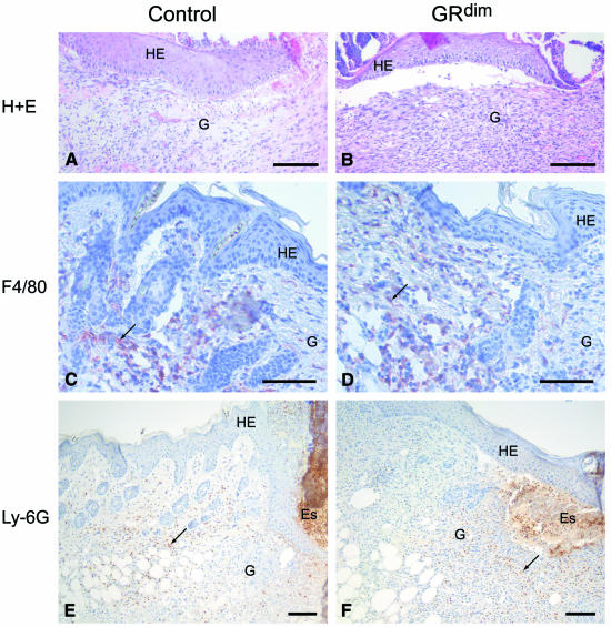
Fig. 2. Granulation tissue composition in 5-day wounds of GRdim mice. (A and B) H/E stainings of the granulation tissue of control (A) and GRdim mice (B) are shown at high magnification. (C–F) Wax sections from control (C, E) and GRdim (D, F) mice were stained with the F4/80 antibody that detects monocytes/macrophages (C, D) or with the Ly-6G antibody that recognizes neutrophils (E, F). See Figure 1 for abbreviations. Scale bars: A–D, 20 µm; E and F, 40 µm.

An official website of the United States government
Here's how you know
Official websites use .gov
A
.gov website belongs to an official
government organization in the United States.
Secure .gov websites use HTTPS
A lock (
) or https:// means you've safely
connected to the .gov website. Share sensitive
information only on official, secure websites.
