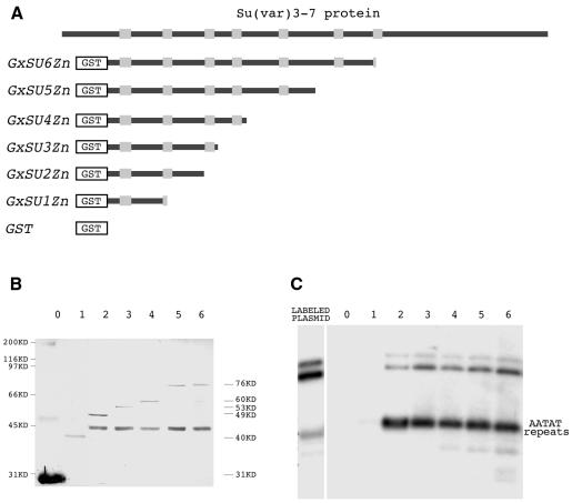
Fig. 2. Binding of the AATAT satellite by different GST–Su(var)3-7 derivatives. (A) The Su(var)3-7 protein is represented as a black bar within which the zinc fingers appear as grey boxes. (B) Western blot of the different recombinant proteins schematized in (A), bound to glutathione–Sepharose 4-B. Lanes 0–6 correspond to proteins with zero to six zinc fingers revealed with anti-GST antibody. (C) Autoradiogram of the agarose gel after electrophoresis of the 300-bp AATAT repeats fragment retained by the same proteins. The amounts of protein fragments in the binding assay were those deposited on the western blot in (B), and the experiment was performed in the presence of 0.1 µg/µl of the non-specific competitor poly(dI–dC).

An official website of the United States government
Here's how you know
Official websites use .gov
A
.gov website belongs to an official
government organization in the United States.
Secure .gov websites use HTTPS
A lock (
) or https:// means you've safely
connected to the .gov website. Share sensitive
information only on official, secure websites.
