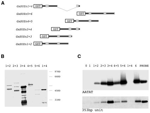
Fig. 3. Binding of the AATAT and 353-bp satellites by different zinc finger pairs derived from Su(var)3-7. (A) Different parts of the fusion proteins are schematized as in Figure 2. (B) Western blot of the proteins corresponding to (A), after purification on glutathione–Sepharose 4-B, revealed with an anti-GST antibody. Lanes are named according to the pair of fingers present in the fusion proteins. (C) Autoradiography of the agarose gel after electrophoresis of the 300-bp AATAT repeats fragment and the 500-bp insert containing the 353-bp unit bound by the GST-tagged protein constructs. The amounts of protein fragments in the binding assay were those deposited on the western blot in (B), and the experiment was carried out in the presence of 0.1 µg/µl of the non-specific competitor poly(dI–dC).

An official website of the United States government
Here's how you know
Official websites use .gov
A
.gov website belongs to an official
government organization in the United States.
Secure .gov websites use HTTPS
A lock (
) or https:// means you've safely
connected to the .gov website. Share sensitive
information only on official, secure websites.
